Application Spotlight: Femtosecond Laser Texturing of Ti-6Al-4V Surfaces
An open-access study in Surfaces maps how femtosecond laser ablation of Ti-6Al-4V evolves from single-pulse craters to scanned areas and finally to a controlled bi-sinusoidal micro-texture. The authors frame the work for dental and orthopedic implant surfaces, where literature links micro-texture to early biological response and osseointegration. This paper itself reports no cell, tissue, or implant testing.
What the Study Did
The team polished Ti-6Al-4V cylinders to Sa about 12 nm, then textured them with an ytterbium fiber laser (Satsuma HP3) at 1031 nm and 306 fs. The beam was linearly polarized with a Gaussian spot of nominal 2w0 = 24.8 µm (M2 = 1.14). Motion used a motorized XY stage with z-axis focal adjustment, not a galvanometer scanner. Surfaces were measured by white-light interferometry in PSI and VSI modes, plus SEM.
The campaign had three steps: fixed-beam single-pulse and multi-pulse craters; scanned 2D areas that varied fluence, overlap, and number of passes; and a 3D bi-sinusoidal pattern with a defined period and peak-to-valley height.
From Threshold to Texture
- Ablation threshold: the single-pulse threshold was 0.175 J/cm2, with no polarization dependence. The incubation factor was S = 0.84.
- 1D crater growth: with a fixed beam, crater diameter grew from about 7 to 32 µm as fluence rose from 0.26 to 6.52 J/cm2. At 0.89 J/cm2, diameter stabilized after about 25 pulses. Depth rose from about 2 µm after 50 pulses to 22 µm after 500 pulses, quasi-linear with pulse number.
- 2D scanning: depth increased roughly linearly with the number of passes up to 90. Roughness and depth depended on overlap and fluence with a non-monotonic trend. An overlap of 50% was selected as a compromise. Sa peaked at 0.23 µm at 60% overlap and 0.49 J/cm2.
Building the Bi-Sinusoidal Surface
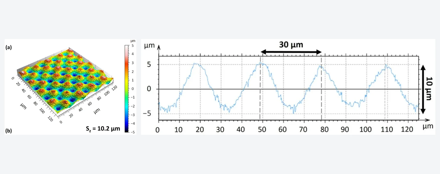
For the 3D pattern the authors used fluence 1 J/cm2, 50% spot overlap, 50% hatch overlap, scanning speed 50 mm/s, repetition rate 5.88 kHz, a slicing step of 100 nm, and four scan orientations at 0, 90, 180, and 270 degrees.
- Period: 30 µm.
- Peak-to-valley height: 10.2 µm before filtering and 9.2 µm after filtering (Sz = 9.2 µm filtered).
- Superimposed roughness: Sa = 120 nm on top of the designed wave.
Why This Matters
The study shows that fluence, overlap, and number of passes can steer metal surface morphology from simple craters to a designed bi-sinusoidal texture with a stated period and height. That helps when a metal surface needs repeatable topography rather than an uncontrolled ablative finish.
The authors' target application is dental and orthopedic implant surfaces. Broader literature discusses micro-texture as a factor in early biological response and osseointegration. Those benefits are not demonstrated here; they are the application context that motivates the process work.
Practical Watch-Outs
- Metrology matters: results are sensitive to measurement and evaluation conditions, and the measured thresholds differ from many literature values, so comparisons require matching methods.
- Non-monotonic roughness: roughness versus overlap and fluence does not rise steadily, so map the process window rather than assuming more overlap gives a smoother surface.
- High pass counts: deviations at high pass numbers can arise from cumulative effects, redeposition, and evolving surface state.
- Optics and motion differ: the study used a stage-based setup with a 24.8 µm spot. Fluence and overlap must be recalibrated for other focusing optics and scanners.
- No biological validation here: the paper does not test cells, tissue, or implants, so implant-surface benefits remain an intended application rather than a result of this study.
Related Clear Photon Pages
For adjacent process context, see Functional Surface Structuring, Ablation, and MedTech & Medical. If you already have an ultrafast source and want a Class 1 workstation path, start with BYOL Laser Integration or Process Development.
Source: Alzarif, H.; Robache, F.; Vayron, R.; Bigerelle, M.; Montagne, A. "Femtosecond Laser Texturing of Ti-6Al-4V: From Ablation Behavior to Controlled Bi-Sinusoidal Surface Morphology." Surfaces 2026, 9(3), 64. DOI: 10.3390/surfaces9030064. Open access under CC BY 4.0. The hero figure is reproduced from Figure 17 of Alzarif et al., Surfaces 2026, under CC BY 4.0; panels are placed side by side for web delivery, with resizing and WebP/JPEG conversion and no other content changes.
Interested in This Application?
Contact us to learn how Clear Photon workstations can enable your research or production process.
Get in Touch